More sophisticated insights into the human mind
“We finally have tools to non-invasively study the human brain in normal subjects and diseased patients,” says Professor Stefan Sunaert, Head of Translational MRI at the Department of Imaging & Pathology at Leuven University Hospital (Belgium).
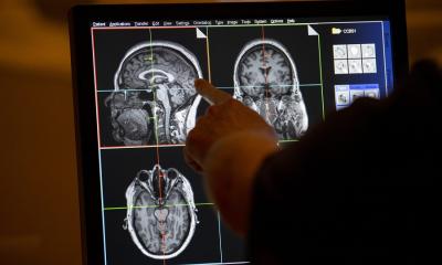
The technology that enables us to perform such path-breaking studies is functional magnetic resonance imaging (fMRI), or to be more precise: resting fMRI (rfMRI), the most recent further development of fMRI which visualizes brain function. rfMRI can show and quantify the brain regions that are involved in a certain process. “But how the brain works exactly is something we still need to discover and learn in order to understand disease,” Sunaert adds. The expert is chairman of the New Horizons Session “Imaging of the mind” at the European Congress of Radiology (ECR 2013), which will end on Monday.
Resting brain activity is identified by looking at changes in brain perfusion. The magnetic resonance imaging procedure uses the different degrees of magnetization of oxygen-poor venous and oxygen-rich arterial blood to create a so-called blood oxygen level dependent signal (BOLD). Based on this signal, brain activity can be visualized and displayed in a color code. Every activity of the brain is associated with a BOLD signal. Thus, the functional organization of a brain and its changes caused by disease can be examined. Unfortunately, the signal is buried under noise so to speak. “It took us a long time to prove that the noise is not only artefacts but that it covers the BOLD signal which indeed correlates to certain brain activities,” Sunaert explains.
The research on resting-state functional connectivity unearthed a number of networks, of patterns of synchronous activities in the brain: the executive control component, the sensory/motor component, the auditory component, up to three different visual components, two lateralized frontal/parietal components and the temporal/parietal component. The elements of these networks are located in different brain regions and in a healthy person they are always simultaneously active.
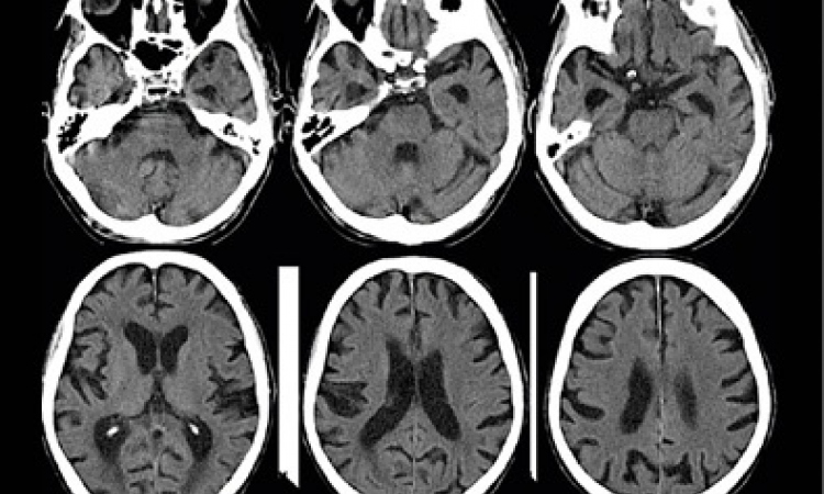
A network that is already particularly well understood is the default mode network (DMN), a web of brain regions that becomes active when a person is awake but in a state of calm. It is activated by reflection, remembering or day-dreaming and it correlates negatively with other networks which are involved in visual perception of the outside world.
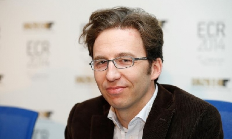
The DMN activity changes over time. “With age the network connectivity decreases. And that is also what happens in Alzheimer’s disease,” points out Professor Andrea Falini, Head of Neuroradiology at the Istituto Scientifico San Raffaele in Segrate near Milano (Italy). Falini’s research focuses inter alia on the differences between natural ageing processes and the development of Alzheimer’s – which all occur in the DMN – with the aim to be able to diagnose Alzheimer’s diseases early. “Early diagnosis is mandatory for new therapeutic strategies,” the Italian neuroradiologist underlines.
Some neuroradiologists are apprehensive about possible abuses of rfMRI. At ECR chairman Sunaert mentioned several applications he personally finds very disconcerting: In one fMRI-based study the neural responses of children in connection with markers were examined. Results confirmed that food logos activate some brain regions in children known to be associated with motivation. Sunaert also pointed out that a US company claims to be able to use BOLD signals to tell lies from truth, a modern version of the lie detector. “That is really scary,” Sunaert shuddered and asked the audience: “Would you undergo an fMRI exam if the technology were able to identify lies or erotic thoughts?” None of the participants raised a hand.
12.03.2013