Article • Trends
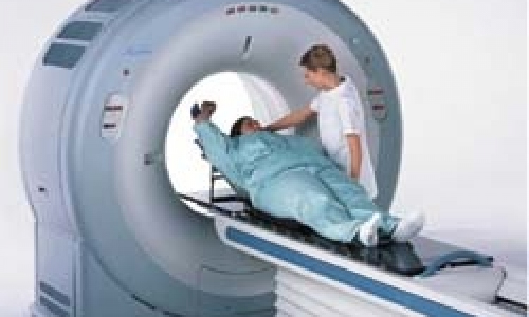
Dose reduction strategies in cardiac CT
World-renowned cardiologists reviewed the latest trends and dose reduction strategies in cardiac CT during the International Conference on Nuclear Cardiology and Cardiac CT (ICNC) that unfolded in Madrid in May.
Report: Mélisande Rouger
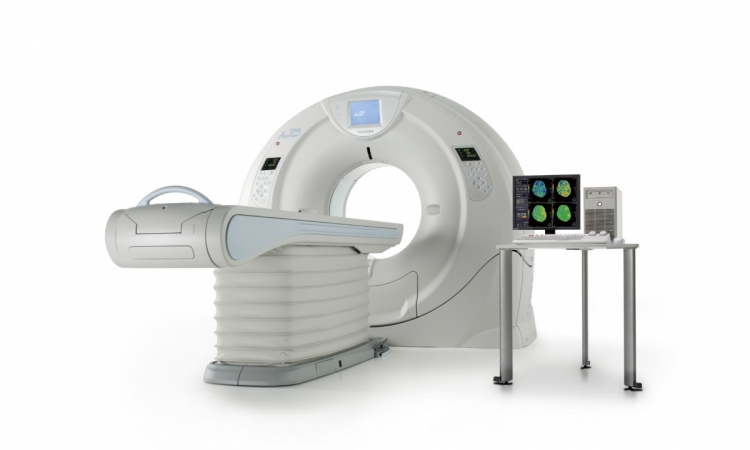
During this year’s International Conference on Nuclear Cardiology and Cardiac CT, Dr Marcio Bittencourt, from Sao Paulo, Brazil, offered an overview of the newest available technology, namely GE Healthcare’s Revolution, Siemens Force, Toshiba’s Aquilion ONE ViSION, and Philips’ Brilliance and IQon Spectral Detector CT scanners.
New scanners must do four things: improve image quality, acquisition speed and coverage, and reduce radiation dose, Bittencourt explained. Temporal resolution – the time needed to acquire one image – should be <15% of the cardiac cycle to minimise motion artefacts. Thus, acquisition time, a challenge in the cardiac setting, must be as low as possible. Faster rotation is one way to achieve that, and most new scanners have indeed increased speed up to 0.25s per rotation. Other options are dual source CT and multi segment reconstruction.
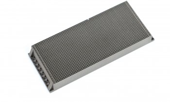
To improve spatial resolution, users can either do sharper reconstruction, although some recent changes in detector technology and flying or dynamic focus spot have also improved spatial resolution. For z-axis coverage, cardiac imaging usually required about 14 cm. Some new scanners now allow this to be performed in a single heartbeat, though this technology is not available for all vendors, Bittencourt pointed out.
New technology enables selection of the best scan mode and protocol for each individual examination, which contributes to reducing radiation dose. Besides protocols, other features, such as automated exposure control, reduced target noise and iterative reconstruction, may also lower dose significantly. One recent technology, spectral energy imaging, has the potential to do calcium subtraction, myocardial perfusion or iodine map, and beam-hardening correction for perfusion.
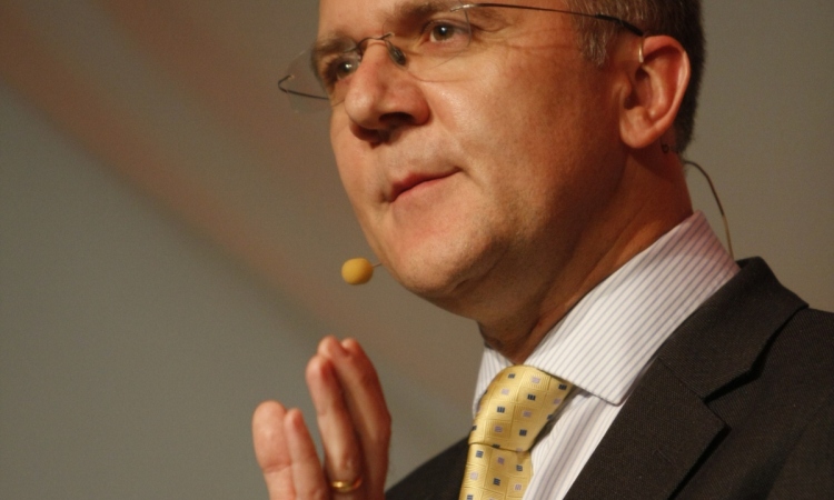
However, not all these options are necessary if users are not doing top-notch research, Bittencourt believes. ‘If you can’t afford newer technologies, any 64 detector scanner allows adequate diagnostic image quality for most patients. Anything newer will cost more. If you ask me whether any of the new scanners better, I think they certainly have improved temporal resolution and spatial resolution, which are interesting and may allow evaluation of more complex patients. So, if you can pay for these new toys, my answer is yes, they are better. But if you ask if they are a cost effective replacement for a 64 detector scanner, from a health perspective, the answer is probably no.’ Dr Stephan Achenbach from Erlangen, Germany, focused on methods for low-dose coronary CTA. ‘CT made its way into European guidelines on stable coronary disease and acute coronary syndrome, so it should really be considered in patient management,’ he said.
There is tremendous potential for dose reduction. A 2007study at 50 sites across Europe compared 1,965 CTA examinations in 2,000 individuals. It showed tremendous differences in estimated radiation dose associated with CT angiography, with some sites using doses of up to 13 mSv on average and others 4.6 mSv. Image quality, however, did not correlate to dose. ‘This study from the past clearly shows that radiation dose can be lowered without sacrificing image quality, and today we have many more options to do so,’ Achenbach said.
The first strategy to limit exposure is to modify the mode of acquisition and to avoid spiral or helical scanning with continuous radiation exposure, which results in a dose in the 25-30 mSv range. ‘That is really inappropriate for most patients who undergo CTA and can easily be modified because, in most cases, we want image reconstruction only in diastole. Most technology enables limitation of the full output of the X-ray tube during the diastolic segment of the cardiac cycle, thanks to ECG-correlated tube current modulation, often called ECG pulsing,’ he explained. Achenbach recommends using ECG pulsing systematically when spiral/helical acquisition is performed, as this will lead to a dose reduction of 40 to 50%.
Prospectively ECG triggered acquisition avoids spiral acquisition and combines step-wise table movements with short periods of data acquisition, typically in diastole. Therefore the dose is low, between 3 and 5 mSv. High-pitch spiral acquisition, sometimes called Flash mode, is a combination of spiral l acquisitions and prospective ECG triggering. Thhis is only possible with dual source scanners and spends low dose, between 1.5 and 2 mSv. However, it requires low and very regular heart rates.
Lowering tube voltage also helps to reduce dose. Traditionally 120 kV were used in cardiac CT, but in many cases this can be lowered to 100 kV. Doing so will reduce the dose by 40%, even in patients who have high body mass index (BMI), according to Achenbach. ‘100 kV should be used in patients less than 85 to 100 kg – some say with BMI < 30 or 25, some combine the two, there are no strict guidelines,’ he pointed out. By combining 100 kV tube voltage with prospectively ECG triggered axial acquisition, dose can be lowered to 2-3 mSv, and to as little as 0.9 mSv with high-pitch acquisition. 80 kVp work in very thin patients (<70 kg), and can lower dose to 0.6 mSv.
Some studies have combined all possible modes for dose reduction and performed coronary CTA with doses as low as 0.1 mSv. However, image quality can be seriously hampered in such an approach. ‘Very low doses are possible, but I have to say I am not a fan for continuing this race for lower doses because we really risk sacrificing image quality and makinf misdiagnosis if we put too much weight on dose. Cardiac CT imaging is not a race to achieve the lowest possible dose; you always have to make sure you retain image quality to evaluate even those patients who have complex situations such as calcified plaque, etc.
Profiles:
Professor Stephan Achenbach is Chairman of the Department of Cardiology at the University of Erlangen, Germany and Vice President of Global Affairs and Communication at the European Society of Cardiology (2014-2016). With major clinical interests in cardiac CT, imaging of atherosclerosis and interventional cardiology, he was president of the Society of Cardiovascular Computed Tomography between 2007 and 2009, and is currently its secretary. He is also a fellow of the European Society of Cardiology, the American College of Cardiology and the Society of Cardiovascular Computed Tomography, and a member of the European Academy of Sciences and Arts.
Dr Marcio Sommer Bittencourt is Assistant Physician at the Division of Internal Medicine, University Hospital of Sao Paulo, Brazil, where he obtained his PhD in 2014. He also gained a Masters Degree in Public Health from Harvard Medical School in 2013, and carried out a post-doctoral research fellowship in cardiovascular imaging at Brigham. His main clinical interests lie in cardiovascular disease, epidemiology, internal medicine, public health, bio- statistics, medical and biomedical image processing and cardiac MRI. He is one of the Fellow and Resident Leaders of the Society of Cardiovascular Computer Tomography SCCT and has over 100 publications to his name. Dr Bittencourt obtained a Masters in Public Health at Harvard Medical School in 2013, and gained his PhD in cardiology from Sao Paulo University in 2014.
28.08.2015