Diagnostic
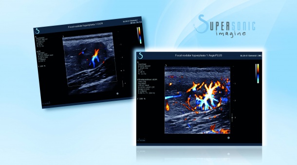
SuperSonic reveals micro vessels with AngioPLUS
Building on an innovative ultrasound technology that continues to yield break-through capabilities, SuperSonic Imagine is introducing AngioPLUS, a third diagnostic functionality for its Aixplorer platform that promises to be instrumental in the diagnosis of cancerous tissues as well as musculoskeletal pathologies.
Previewed in October, 2015 during the French Radiology Congress, AngioPLUS will begin shipping on the Aixplorer after its global launch at the annual meeting of the Radiological Society of North America. "This innovation is possible thanks to the UltraFast acquisition and plane wave imaging we applied first to ShearWave Elastography for the real-time assessment of lesions, and that we extended by introducing UltraFast Doppler last year," said Jacques Souquet, Chief Innovation Officer of SuperSonic Imagine, and the company's co-founder.
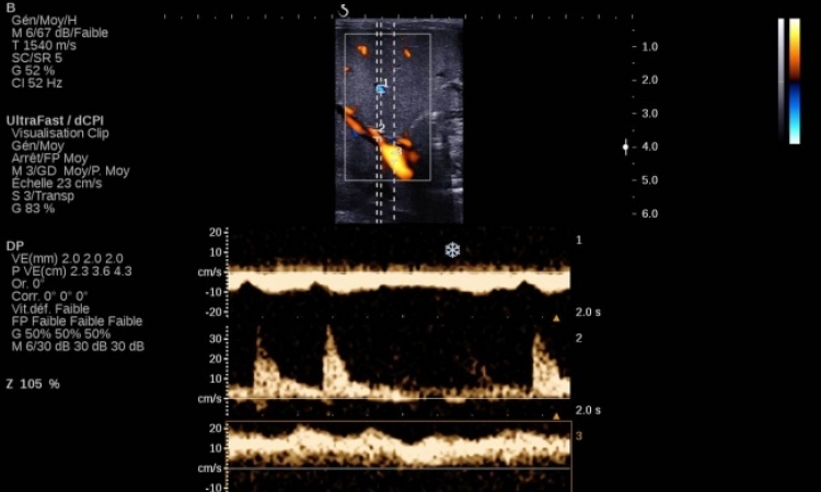
AngioPLUS leverages the capabilities of UltraFast Doppler to provide a new level of microvascular imaging for vascular evaluation. Color sensitivity and spatial resolution are significantly improved and when combined with exceptional 2D imaging, the result is an increased detail of real-time flow information available during ultrasound diagnostic exams. "We have improved sensitivity, compared to conventional Doppler, by a factor of 50 and this enables us to look at the microvasculature of lesions, the angiogenesis of tumors, and potentially without the use of contrast agents," said Souquet.
According to Souquet, one of the frustrations for clinicians is that with conventional Doppler they can see the beginning of the slower flow that indicates micro vessels, but they are unable to extract the image. "It is something that they simply can not see without enhancing the ultrasound signal using a contrast injection, and what AngioPLUS can do is to enhance that signal without contrast," he said. Currently the only technique for seeing micro vessels is to illuminate them with an injection of micro air bubbles into the bloodstream.
AngioPLUS may reduce the need for interrupting exams of micro vessels in order to inject a contrast agent, Souquet said, and become instrumental in aiding the diagnosis of cancerous tissues in areas such as the breast, liver, lymph nodes and thyroid as well as musculoskeletal pathologies such as inflammation in tendons."The advantage of out technology is the ability to use plane wave imaging on the Aixplorer. Using conventionally acquired Doppler would require a lot of filtering of their signal, looking at images sequentially, and trading off either frame rate or performance," said Souquet. "Thanks to the UltraFast acquisition, we don't need to make this kind of compromise, and going even further, with our platform what we can do is provide a quantification of things like flow direction."
According to Prof. Jean-Michel Correas, Vice Chairman of the Adult Radiology Department, Necker University Hospital, Paris. "The AngioPLUS technology significantly improves flow sensitivity in color imaging. This innovation allows physicians to quickly and accurately address challenging clinical situations such as characterizing fortuitously discovered focal liver lesions and renal blood flow disturbance. We believe AngioPLUS can help avoid additional imaging or biopsy procedures." With the Aixplorer ultrasound platform, SuperSonic introduced a disruptive technology for ultrasound by swapping conventional line processors for a graphics processor used by the video gaming industry to create lightning-fast sonic image acquisitions.
ShearWave Elastography was the leading edge for the company to differentiate itself in the crowded ultrasound space. Progressively the company demonstrated in more than 60 peer-reviewed publications, including a multinational study of over 1,600 patients the benefits of using the ShearWave technology for the diagnosis of breast lesions. Results showed that ShearWave Elastography, combined with conventional ultrasound criteria, allowed superior accuracy in the diagnosis of breast lesions, significantly reduced the number of false positive cases and helped reduce the number of negative biopsies.
More recently the company announced the enrollment of 2,270 patients for a milestone multi-center study conducted in 22 locations across China to analyze the benefit of combining ShearWave evaluation of individual breast lesions to their classification using the Breast Imaging Reporting and Data System (BI-RADS) by the American College of Radiology. In addition to breast cancer management clinical studies, over 60 liver disease-focused publications have demonstrated the reliability and effectiveness of ShearWave Elastography to assess the severity of chronic liver disease. In April 2015, results of a 1,340 patient study confirmed the accuracy of ShearWave as a non-invasive alternative to biopsy for staging liver fibrosis.
01.12.2015