Sodium
How ‘salt’ MRI scans could give a clearer picture of disease
A novel technique to use the body’s natural sodium (salt) content to provide a more detailed picture of tissue health and disease is to be pioneered by MRI experts at The University of Nottingham.

The world-leading team at the Sir Peter Mansfield Imaging Centre (SPMIC) has been awarded just over £1m to develop the untapped potential of Sodium MRI as an advanced scanning technology.
Current clinical MRI uses hydrogen in the body’s water and fat to produce scans, but this does not provide all the information about tissue health and disease progression stages. Sodium ions naturally occurring in the body are much smaller than water molecules and are involved in many body functions associated with pathology. Sodium MRI has great potential to be a useful new high and ultra-high field scanning target in the future.
Kidney disease will be the main application of the research working in collaboration with the Centre for Kidney Research and Innovation (CKRI), but the team believes that Sodium MRI can also be used for more accurate diagnosis and monitoring of other diseases, and perhaps will give new insights into disease mechanisms as sodium
management is important in the brain, lung, liver, and musculoskeletal system.
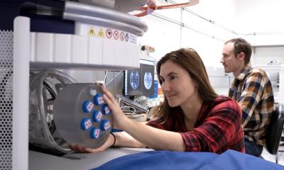
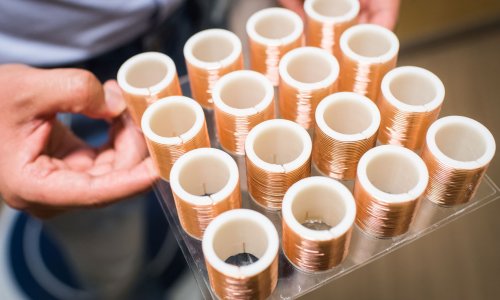
The MRC Discovery Grant of £672,000 with a £369,000 investment from the University will allow the researchers to develop sodium MRI on the 3T and 7T magnetic field scanners at the SPMIC, University Park Campus, Nottingham. The team will use MRI coils for sodium imaging, these are the receivers of radiofrequency signals in the MRI scanner specifically tuned to the resonant frequency of sodium ions. The researchers will develop new pulse sequences so these new coils can image the torso and limb. The team hopes eventually to take Sodium MRI technology from bespoke research into real world healthcare in healthy volunteers and patients.
Translational imaging researcher Dr Galina Pavlovskaya, School of Medicine, SPMIC said: “Sodium MRI is a novel and undeveloped technology which has huge potential for the healthcare of the future. We are delighted to receive the funding to take it forward at Nottingham. The technique of using sodium ions in the body as a biomarker for imaging is very challenging because of the lower detectability of the sodium signal in biological tissue by currently available MRI scanners. However, high and ultra-high magnetic field scanners available in the Centre should be able to help to circumvent this obstacle.
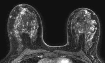
”Sodium ions are much smaller than the hydrogen protons bound to oxygen molecules in the water in our bodies which are mapped by conventional MRI. Therefore, sodium has the capacity to give us a much clearer and detailed picture of the structure and health of an organ from deep inside the tissue. Our aim is to refine the technology so we can turn theory into clinical reality.”
Co-researcher Dr Susan Francis, School of Physics, SPMIC added: “The team has a special interest in new types of functional MRI using novel targets like sodium as quantitative biomarkers of disease in the body, in particular in the kidney. The kidney is an ideal target for our project because it is important in the regulation of sodium in the body. If we can image how sodium is distributed in the kidney and how that differs in a diseased kidney, the impact on the diagnosis and treatment of kidney injury or disease is potentially great. The technique also has specific relevance to understanding sodium and water balance in dialysis patients.
“Sodium accumulation in the skin is a biomarker for kidney stress but we will also be examining how salt is distributed in the skin and muscle. This has lots of clinical applications, for diseases like muscular dystrophy for example. Theoretically we should also be able to study sodium concentration in red blood cells which again has major implications for disease diagnosis and treatment. The technique will give a picture of the body’s naturally occurring sodium and how it is trafficked in the body and will not require any invasive addition of sodium to the scanned subject.”
The work will involve scanning healthy volunteers, and combining conventional Proton MRI to track water in the body with Sodium MRI to analyse the concentration and flow of sodium ions. Chronic kidney disease or acute kidney injury patients will then take part in a second small trial to assess the new Sodium MRI technique.
The project will end in 2018 with a Sodium MRI Conference for the worldwide MRI research and clinical community.
Source: University of Nottingham
18.03.2016