Sponsored • The IQon Spectral
Accustomed workflow, low dose and more precise diagnostics
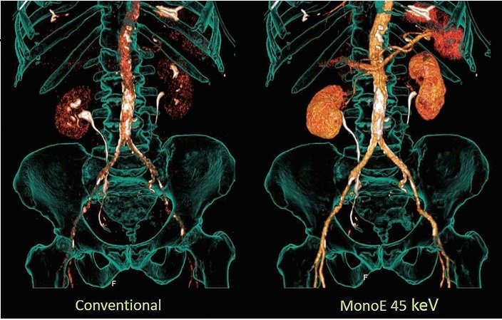
For the purposes of additional diagnostic information, spectral imaging utilizes the effect that the weakness and absorption of x-rays are dependent on their energy level and the tissue through which the radiation passes. Herein the specific and quantitative detection of iodine contrast agent is of particular interest. For example, it makes it possible to calculate out the entire background of a region, including bones. The radiologist obtains a better overview of the conditions, significantly improving the determination of findings.
Examples of using the method
In peripheral arterial occlusive disease, the person making the findings can more easily recognize and assess the vascular tree. In intracranial aneurysms and arterio-venous malformations, three-dimensional reconstructions and projection images without bones considerably speed up the assessment. Last but not least, speed is a major advantage when diagnosing stroke.
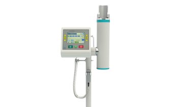
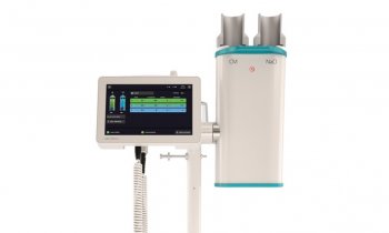
The IQon Spectral CT from Philips
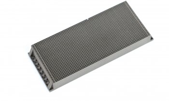
The IQon Spectral CT from Philips – a completely new development based on Dual Layer detector technology – has the ability to detect structures based on tissue composition, and to better differentiate and characterize them. With the Dual Layer detector, which can differentiate between x-ray photons at different high and low energy levels, the IQon Spectral CT opens up new dimensions in CT imaging. The Dual Layer detector design allows for perfectly aligned acquisition in time and spatial terms. This results in data that can be fully reconstructed in projection domain, generating unique quantifiable Spectral information. As a result, eg, precise mono-energetic images can be reconstructed with low noise across the entire range from 40keV to 200keV. Having this at your fingertips offers great value of enhancing Iodine or reducing artifacts.
Use also retrospectively
Another advantage of the Dual Layer detector design is that in the IQon Spectral CT, spectral information is obtained in parallel during normal image acquisition. Aside from conventional images, every 120kV scan simultaneously provides Dual Energy data sets – even if the clinical inquiry does not initially indicate it. In other words: The radiologist no longer has to make the decision as to whether use a normal CT image or a spectral protocol before the scan. Both data sets are available after the examination. If suspicious structures or structures which are difficult to interpret are observed in the “normal” CT scans, additional information about the tissue composition becomes desirable, the system can immediately provide the spectral information. If required, each scan can also be shown spectrally without added time requirements due to a second scan and without the additional dose required for a second scan. Users have the security of being able to fall back on a broad range of Spectral information like virtual MonoE and VNC images, using the MagicGlass Window technique as needed.
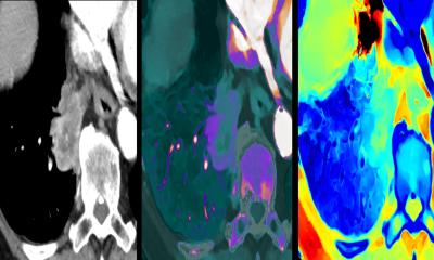
The MagicGlass tool simultaneously shows spectral data in colour if one guides it over the underlying HU image dataset (HU = Hounsfield units). Whether there is an additional need for tissue characterization, contrast enhancement or artifact reduction, the information is there to provide the support for the diagnosis and make diagnostics more convenient.
04.05.2016