Memory
A study shows how the brain switches into memory mode
Researchers from Germany and the USA have identified an important mechanism with which memory switches from recall to memorization mode. The study may shed new light on the cellular causes of dementia. The work was directed by the University of Bonn and the German Center for Neurodegenerative Diseases (DZNE).
Because of its shape, the control center of memory bears the poetic name of “hippocampus” (seahorse). New sensations to be stored continually enter this region of the brain. But at the same time, the hippocampus is also the guardian of memories: It retrieves stored information from the depths of memory.
The hippocampus is also an important transport junction. And just like rush hour in a major city, it also needs a regulating hand to control the opposing flows of information. The researchers from Bonn, Los Angeles and Palo Alto have now identified such a memory traffic policeman. Certain cells in the brain, the hippocampal astrocytes, ensure that the new information is given priority. The mind thus switches into memorization mode; by contrast, the already saved memories must wait.
However, the astrocytes themselves only take orders: They react to the neurotransmitter acetylcholine, which is released in particular in novel situations. It has been known for several years that acetylcholine promotes the storage of new information. How this happens has only been partly understood. “In our work, we were able to show for the first time that acetylcholine stimulates astrocytes which then are induced to release the transmitter glutamate,” explains Milan Pabst, who is a doctoral candidate at the Laboratory for Experimental Epileptology of the University of Bonn. “The released glutamate then activates inhibitory nerve cells which inhibit a pathways mediating the retrieval of memories.”
The researchers working with the neuroscientist Prof. Dr. Heinz Beck genetically modified nerve cells so that they could be activated by light and then release acetylcholine. Using this trick, they were able to clarify the mechanism using recordings in living brain tissue sections. “However, we also show that, in the brains of living mice, acetylcholine has the same effect on the activity of the neurons,” explains Pabst’s colleague, Dr. Holger Dannenberg.
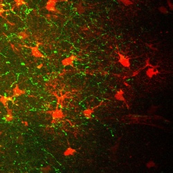
Astrocytes have long since been underestimated
Another reason this result is interesting is because astrocytes themselves are not nerve cells. They belong to what are known as glial cells. Until the turn of the millennium, they were still considered to merely serve as mechanical support to the real stars of the brain, the neurons.
In recent decades, however, it has become increasingly clearer that this image is far from correct. It is known by now that astrocytes can release neurotransmitters – the messengers by which neurons communicate with each other – or even remove them from the brain. “It was previously unknown that the astrocytes are involved in central memory processes through the mechanism which has now been discovered,” explains Prof. Beck. However, an observation made by US scientists in 2014 fits into this context: If astrocytes’ function is inhibited, this has a negative effect on the recognition of objects.
The results may also shed new light on the cellular causes of memory disorders. Thus there are indications that the controlled secretion of acetylcholine is disrupted in patients with Alzheimer’s dementia. “However, we have not investigated whether the mechanism we discovered is also impacted,” stresses Pabst.
Source: Rheinische Friedrich-Wilhelms-Universität Bonn
06.05.2016