Article • Mini Microscopes
Mobile technology could enhance digital pathology
Mobile technology could prove an important catalyst in helping take digital pathology onto a new level in delivering clinical diagnostics.
Report: Mark Nicholls
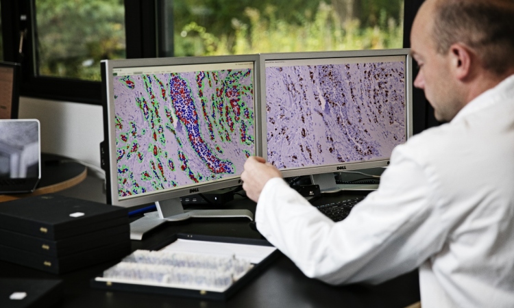
For a number of years, pathology has moved more slowly than other fields that have embraced digitisation, primarily because of the costs and scale of the required equipment, according to Professor Johan Lundin, who is Research Director at the Institute for Molecular Medicine Finland (FIMM), hosted by the University of Helsinki. An inability to take digital microscopy out of the laboratory as a portable tool has also been a consideration, however, that is about to change with a number of mobile digital microscopes in development.
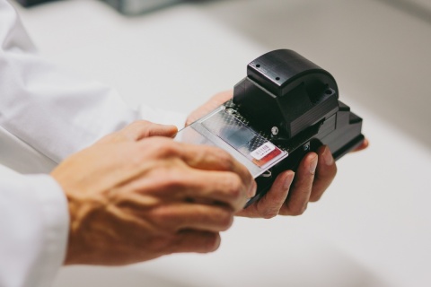
Among them is Professor Lundin’s research group at FIMM and Karolinska Institutet in Stockholm, which has been working to pioneer the MoMic portable scanner, currently undergoing tests in Finland and Tanzania. He explained: “Having the microscope closer to patient, or where the samples are obtained, would improve the way you can perform diagnostics, making it more rapid and more accessible. It would improve the workflow of diagnostics, not just in pathology but also microbiology, cytology and forensic medicine.”
The low-cost, easily transported mobile microscope and scanner developed by the Finnish researchers enabling the use of rapid, computer-assisted remote diagnostics, uses cheap components originally intended for mobile phones and connects wirelessly to a cloud server to which it transmits images for machine vision analysis.
“Digital pathology, and digital microbiology, can make much use of technology from the mobile phone industry if combined with the optical components you need for microscopy,” added Professor Lundin, who is chairing sessions at the 13th European Congress on Digital Pathology in Berlin from May 25-28.
MoMic is still an academic study, but what differentiates it from similar mobile microscopy projects is the scanner facility it incorporates, which is vital as it enables the digitisation of the whole sample. “If you cannot digitise the whole sample it is very difficult to use it for medical diagnostics because it is extremely difficult to control where the important part of the sample is on the slide,” he pointed out.
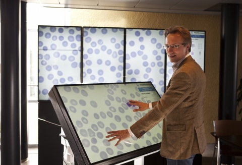
The advantage of MoMic is that it becomes a point-of-care device and can attain laboratory-levels of microscopic resolution and scan a sufficient area of the sample to enable diagnosis. Once a sample is obtained it can facilitate faster digital diagnosis or the ability to consult remotely.
Professor Lundin said the MoMic project started with the goal of creating an instrument for low resource settings and currently five prototypes are being tested. Field studies are under way in a small hospital in a provincial area of Tanzania, a country where there are less than 20 pathologists among a population of 45 million people.
In the three-year pilot programme, patient samples will be diagnosed both on location and remotely based on the digitised sample. In such countries, he believes MoMic will make an impact in the diagnosis of cancer as well as infectious diseases including malaria, tuberculosis or stool parasites. Other tests are under way in Finland for cancer diagnostics during surgery, using the device away from the lab for pathologists to view samples to check tumour margins and ensure the lymph nodes are clear from cancer.
The next steps, supported by a government innovation grant, are to establish routes to commercialisation with partners having been identified to transform it into a product from the prototypes, with large-scale validation studies planned for next year.
Professor Lundin also sees portable digital pathology microscopy being used in parallel with larger high throughput scanners, supporting single pathologists in smaller units, a move he believes would open the world of digital pathology to more practitioners. The mobile microscope would also be well suited for research work, including the study of tissue and cell samples to identify the expression of biomarkers in cancer research, believes Professor Lundin.
Profile:
Professor Johan Lundin is Research Director at the Institute for Molecular Medicine Finland (FIMM) hosted by the University of Helsinki and Guest Professor in Medical Technology at Karolinska Institutet, Stockholm, and also Associate Professor in Biomedical Informatics at the University of Helsinki. His key research aims are to fully utilize development within information and communication technologies for improvement of diagnostics and care of the individual patient.
23.05.2016