'More knowledge — less dose'
By Guido Gebhardt
X-rays are made up of high-energy photons. This type of radiation follows the physical laws of electromagnetic waves as well as of particles.
Just like the filament in a light bulb a hot-cathode emits electrons. These are subjected to high speed acceleration toward the anode by an electric field. When the electrons penetrate into the anode disk upon impact they undergo severe deceleration. The energy thus released is emitted as X-ray radiation. The high voltage applied is a measure of the energy; the tube current – the number of electrons being accelerated toward the anode – controls the intensity. When both parameters are well-adjusted the radiation exposure of the patient is minimised.
The word ‘dose’ describes the energy effect an X-ray transfers to the material. Therefore, the radiation dose is quantified in terms of the amount of energy (unit: Joule) transferred by radiation to a certain amount of material (unit: kilogram). In this context the dose is known as absorbed dose. The unit of absorbed dose is Gray – abbreviated as Gy. One Gray is equal to one joule per kilogram (1Gy = 1 J/kg).
Different tissues absorb radiation to different degrees. The amount of radiation absorbed by the body is also expressed as a dose. Different types of radiation, e.g., alpha radiation, neutron radiation and x-rays, trigger different relative biological effects in the tissues, which scientists account for by assigning weighting factors to them. The product of absorbed dose and the weighting factor of the radiation is known as the equivalent dose. Its unit is Sievert, abbreviated as Sv. X-ray radiation has a weighting factor of one and therefore the equivalent dose is equal to the absorbed dose.
Exposure to natural and medical radiation – Human organs differ in their susceptibility to radiation; the skin, for instance, is rather insensitive, while the gonads – in women, ovaries and, in men, testicles – are most susceptible. This is accounted for by the tissue weighting factor. The product of equivalent dose and the tissue weighting factor – summed over all organs irradiated – is known as the effective dose. The effective dose is the usual measure of the radiation exposure of the patient and is also given in Sievert (Sv or mSv for one thousandth of one Sievert).
The impact of the dose/effect of ionising radiation on the DNA in body cells is subject to scientific controversy. It has been demonstrated that ionising radiation can alter the genome. However, experts disagree on which dose affects which degree of damage.
Radiation exposure in CT scans
State-of-the-art CT scanners cause additional radiation exposure of 2–20 mSv, depending on the model and the region of the body studied. Studies confirmed damage to the body only for a dose of at least 0.5 Sv – that is about 500 times the CT radiation exposure. Epilation of the skin becomes a risk only with an absorbed dose of at least 3 Sv (about 1,000 CT studies). Therefore, deterministic radiation damage (damage that can be traced to a certain event) due to CT studies can be ruled out completely. The data on stochastic damage (probability of damage after irradiation) is founded on long-term observation of the survivors of Hiroshima and Nagasaki. Since 1950, the so called life span study, covering about 120,000 patients, has confirmed a linear relationship between dose and additional risk of cancer. To a large extent, this risk depends on the age when irradiation happened. If irradiation took place during childhood the risk is increased. ICRP 1990 (International Commission on Radiological Protections) hypothesises an additional lifetime mortality risk of cancer of about 5% per Sv. In other words, a CT study with 10 mSv carries an additional mortality risk of 0.05%.
Compared with other known risks this is a rather low value. Cardiovascular disorders increase the mortality risk by 34%, heavy smoking by 40% and alcohol abuse even by 72%. If the general risk of cancer is 25%, a CT study will increase this risk to just 25.05%. Without a doubt, for medical indications the patient benefit far outweighs the additional risk of radiation exposure.
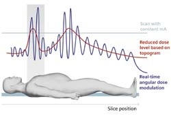
Modern CT scanners are equipped with numerous tools that allow radiologists to keep patient radiation exposure as low as possible. Decreasing patient diameter by just 4 cm will halve the tube current. For quite some time, automatic dose modulation, such as CareDose4D by Siemens, has been taking this into account. It ensures real time automatic dose modulation
by anatomically controlled automatic exposure control, thereby reducing the dose by up to 66%. Asymmetric collimator control preventing over-exposure of the area studied and ECG-gated dose modulation also help to reduce radiation exposure even further. Knowing how to handle the unit properly has a direct impact on patient dosage.
28.10.2008