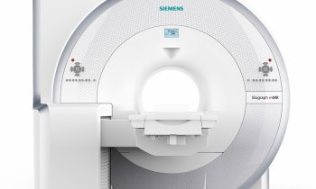
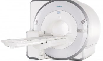
The first whole-body MRI-PET system
The technological integration of positron emission tomography (PET) and magnetic resonance imaging (MRI) has been the dream of molecular imaging experts and engineers for some time. Now, the German Science Council has agreed to provide 6.56 million funding to install a whole-body MRI-PET prototype in the centre of excellence for imaging procedures at the radiology clinic in Eberhard-Karls University, Tübingen, Germany. Construction of the housing for the new system will begin this year, with the equipment scheduled for use in 2011.

The Tübingen working group, headed by the medical director of the university hospital’s radiology clinic, Professor Claus D Claussen (CC) who has worked with colleagues Professor Heinz-Peter Schlemmer, head consulting physician for MRI, and Professor Dr Bernd J Pichler, head of the pre-clinical imaging and imaging technologies laboratory, and Siemens Healthcare in Knoxville, Tennessee, as well as Siemens MR, Erlangen, Germany, to develop an MRI-PET scanner.
‘We believe the MRI-PET hybrid technology to be of particular value to onco-diagnostics and to stroke and cardiac diagnostics,’ said Prof. Claussen. ‘In Tübingen, we have already taken the first steps towards this technology. The first combined equipment, which was developed by Professor Pichler and team, is a 7-tesla MRI-PET scanner for small animals. Since it met with huge interest from research labs, it is now being marketed by a tomography manufacturer for research purposes. The demand, particularly in the pharmaceutical industry, is high because such a scanner allows for non-invasive examinations, for example of mice in bio-medical basic research. Such an MRI-PET scanner is ideally suited to document the course of a disease and therapy success. The encouraging interest in MRI-PET imaging technology by industry and research institutions has been a driving force for our work. From early 2008, we have been testing an MRI-PET hybrid system developed by Siemens. This 3-tesla prototype, which is based on the small animal scanner, is used for brain diagnostics. We hope to gain new insights for diseases such as Parkinson’s, epilepsy or Alzheimer, and regarding neurological stem cell therapy. After having improved this highly complex technology in a joint effort with Siemens, we’re about to begin first clinical trials.
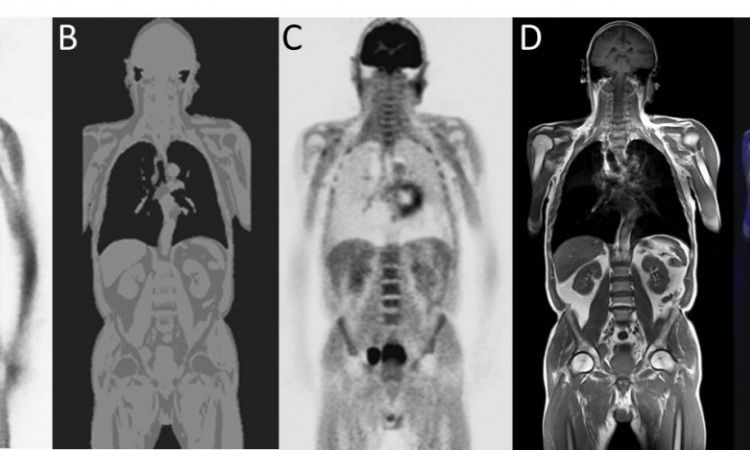
Asked what the MRI-PET advantages are over PET-CT, the professor said that the MRI-PET combination brings a manifold increase of the potential of both modalities. ‘MRI offers much better contrast resolution than CT and can provide metabolic information, for example with MRI spectroscopy. With the help of radio-active tracers, PET on the other hand shows regions with increased metabolic activity in great detail and with high sensitivity. MRI can detect substances on the millimolar (mM) level, PET can do this on the picomolar (pM) level. Thus, a combination of MRI and PET is the ideal synthesis of functional and morphological imaging. A further advantage: it is a hybrid system, which enables simultaneous image acquisition rather the post-acquisition fusion of images, as is the case with PET-CT. This not only reduces examination times but also offers parallel documentation of interacting functional processes, thus providing much more precise information. The examination results are available quicker and they are more exact, so relevant data regarding a specific therapy – or for therapy control purposes – are available sooner. Not to forget radiation exposure: PET-MRI doses are only half of those required by PET-CT scanners because only the tracer involves radio-active substances.’
Facing technological challenges
First and foremost the detectors must be constructed in such a way as to work in a magnetic field, Prof. Claussen pointed out. ‘Conventional PET detectors use photo multipliers, which can be compared to a TV tube whose signals are distorted in a magnetic field. Professor Pichler has been working on MRI-compatible PET detectors for many years. With his team he developed avalanche photo diodes – APDs – which are now used in our head and whole-body scanners. These chips are based on semiconductor technology and are not affected by the MRI system’s magnetic field.
‘A further problem is the implementation of a suitable PET attenuation correction as it is provided by X-rays in a PET-CT scanner. Adequate correction of the radiation attenuation in human tissue in the PET scanner is the precondition for unmarred visualisation of the tracer uptake throughout the entire region of interest. In a conventional PET scanner you use the CT data for the correction of scatter and absorption. This is obviously impossible in an MRI-PET scanner. We therefore developed segmentation algorithms similar to those used in a CT scanner. In a joint development with the Max Planck Institute in Tübingen, we were able to implement this method successfully in head scans. However, in whole-body scans things are more complicated due to complex anatomies and distortions created by breathing. Finding a solution is a highly complex task.
13.11.2009