The ultra-high-field MRI symposium
Early problems of ultra-high field Magnetic Resonance Imaging (MRI) have been overcome by successful development of adequate hardware. In consequence big efforts have been achieved in structural imaging, as well in functional imaging. Basic scientists and physicians who work in ultra-high-field MRI in Europe and the USA, met at the Berlin Ultra-high-field Facility (BUFF), in the Max Dehlbrück Centre (MDC), Germany, to discuss their findings at the 1st annual symposium.
According to the event organiser, Professor Thoralf Niendorf, head of the facility, the main goal of the symposium was to introduce the BUFF to the international UHF-MRI community and to initiate an annual gathering to provide a regular overview of state-of-the-art in UHF MRI, discussions on future directions, foster joint explorations and initiate international collaboration.
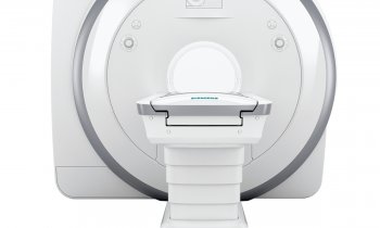
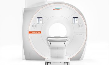
Giving an insight into work at the facility (launched in January 2009, and headed by Prof. Niendorf in August that year), he explained that it possesses a 3-T, 7-T whole-body human MR scanner and 9.4-T animal MR-scanner. Different research teams, which include basic scientists, mathematicians, engineers and clinical scientists, focus on the development of UHF MR applications in cardiovascular, neurological and cancer research.
Working with the 7-T whole body MRI scanner includes development of appropriate hardware. ‘We received a very well designed MR system. Due to the early stage of the explorations there was limited extra ancillary hardware available -- RF-coils suitable for cardiac and body imaging, for example. That’s why we set up an environment where we build MR equipment and improve existing hardware,’ he explained. Several ultra-high-field centres already have developed prototypes of novel MR-coil hardware and are now in a process of having this hardware certified for clinical applications.
Barriers against 7-T going clinical
Dr Franz Schmitt, Director of Ultra-high-field MRI and Collaboration Management in the Siemens AG Healthcare division, the first company delivering a 7-T to Massachusetts General Hospital in 2001, lectured on the remaining barriers for the 7-T to go clinical.
There are still regulatory obstacles. The 7-T is still neither CE certified nor other governing bodies. However, according to Dr Schmitt, in 2003 the FDA agreed to allow UHF MRI up to 7-T, but this is blocked by the regulatory of International Electrotechnical Commission (IEC). The International Commission on Non-Ionising Radiation Protection (ICNIRP), a non-governmental organisation officially recognised by the World Health Organization (WHO), changed its perspective in September 2009 and classified in a guideline the use of up to 8-T as safe in a controlled mode.
A directive of the European Parliament and of the European Council (2004/40/EC), which is supposed to provide measures to protect workers from risks related to electromagnetic fields (EMF) requires special attention for the daily, routine use of UHF MRI in a clinical environment. In this context, the Alliance for MRI was formed in 2007. This body aims to safeguard the future use of MRI through an EU-wide exemption for use in medical and related research from any exposure limit values set in the Physical Agents 2004/40/EC (EMF) Directive. (Details on the Alliance for MRI are published in our Online Edition www.european-hospital.com 01/06/2010) .
Another barrier against 7-T MRI going clinical is currently associated with the large, passively shielded magnets and their overall costs of ownership. Soon, this obstacle will be overcome due to the introduction of actively shielded magnets, Dr Schmitt pointed out. In contrast to the passively shielded magnet, which requires tons of iron for shielding, the actively shielded magnet makes extra iron shielding almost obsolete. For example, a third of the total cost of the 7-T MRI at BUFF was spent on the iron shielding cage that surrounds the magnet.
Ultra-high-field MRI of the brain
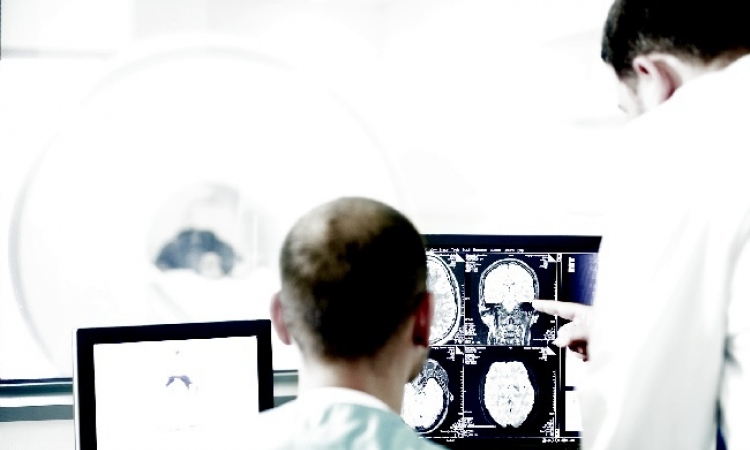
During the meeting, UHF MRI pioneer Professor Lawrence L Wald, member of the A Martinos Centre for Biomedical Imaging in Charlestown, Massachusetts, USA, and associate Biophysicist at Massachusetts General Hospital, presented his results on depth-resolved laminar analyses of resting-state fluctuation amplitude in high-resolution 7-T in functional MRI (fMRI). In the past, high spatial resolution MRI was feasible for structural or anatomical imaging only. Achieving a submillimetre spatial resolution in brain fMRI has been elusive as yet, due to the competing constraints of temporal resolution of 2-3s and image quality. Using the sensitivity advantage of 7-T MRI in conjunction with dedicated, home-built RF coils, Prof. Wald and team have been able to produce functional brain images that show subtle intracortical structures (0.5 mm) using 7-T fMRI.
Professor Arno Villringer, Director of the Department of Neurology at the Max Planck Institute for Human Cognitive and Brain Sciences, Leipzig, Germany, presented studies related to the pathogenesis of obesity in women and possible indicators in the brain. With fMRI a 7-T, he said, it will be possible to detect patterns of activation and deactivation of different sub-areas in the primary somatosensory cortex where, up to now, only a blur of activation can be seen.
Professor Ulrich Dirnagl, Deputy Executive Director of the Centre for Stroke Research at the Charité University Medicine, Berlin, also sets his hope in functional UHF MRI. ‘MR is already in the pole position in the spectral diagnosis in stroke and of course in gives us morphology, together with early markers of tissue damage such as perfusion and diffusion weighted imaging,’ he said, adding that from functional ultra-high-field MRI he expects early tissue signatures, indicating a stroke, as well as more detailed insights into pathophysiological brain changes.
UHF MRI in cardiology
Speaking on 7-T in cardiac imaging, Saskia van Elderen MD, of the Department of Radiology, Leiden University Medical Centre (LUMC), who collaborates with Professor Andrew Webb, said: ‘In a direct comparison with lower field strength 3-T, increased signal-to-noise ratio, improved contrast-to-noise ratio and improved vessel sharpness were obtained at 7-T within a comparable scan duration. The increase in signal-to-noise ratio can be traded for spatial resolution, which is interesting, for example, in vessel wall imaging, visualising positive outward arterial remodelling one of the earliest stages of atherosclerosis.’
Pioneer in cardiac MRI, Professor Jeanette Schulz-Menger of the Charité Medical University, demonstrated that cardiac chamber quantification at 7-T using 2-D CINE FGRE is feasible and agrees closely with LV parameter derived from 2D CINE SSFP imaging at 1.5. The signal gain at 7-T helped to identify fine, subtle anatomic structures, such as pericardium, mitral and tricuspid valves and their associated papillary muscles, and trabeculae.
Tommy Vaughan, of CMMR in Minneapolis, USA, pointed out that 7-T CMR applications become increasingly a research tool, including early explorations into the assessment of regional wall motion using tagging techniques and the assessment of the applicability and efficacy of SSFP imaging techniques at 7-T. According to Prof. Niendorf it should be noted that CMR at 7.0 T is still in its infancy and needs to continue to be very carefully validated against CMR applications established at 1.5-T and 3-T.
The future
In the final session the future perspectives of UHF MRI were assessed.
Outstanding in dimension is the project to build an 11.7 Tesla whole body system at Neurospin, a centre belonging to the Atomic Energy Commission (CEA) in France. Engineer Christopher J Wiggins PhD explained that the aim of NeuroSpin is to push the current limits of MRI and spectroscopy as far as possible in order to study the central nervous system (Further details: www.european-hospital.com, a John Brosky report in our Online-Edition. 11/12/2009).
In his lecture, Professor Mark E Ladd, from the Erwin-Hahn-Institute for Magnetic Resonance Imaging in Essen, asked whether 7-T will go clinical -- and answered his own question: ‘Not yet’. Although technical performance has been demonstrated for particular cases, diagnostic performance preliminary in very small case studies, as yet there has been no demonstration of diagnostic or therapeutic impact, patient outcome, or societal efficacy.
16.07.2010