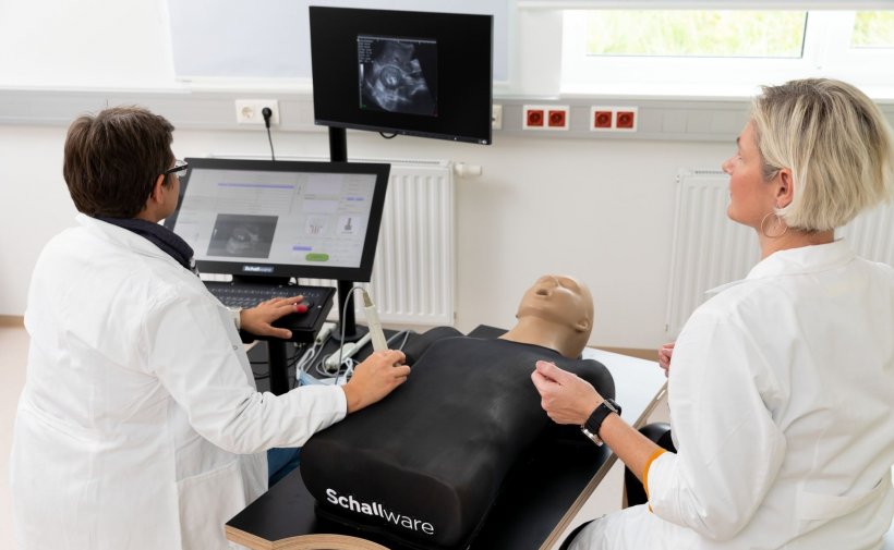
© FH Campus Wien/Ludwig Schedl
Article • Sonography challenges
Abdominal ultrasound: promising for imaging of obese patients
Imagers need special scenarios, skills and tools to examine larger patients, who present more challenges when it comes to radiological procedures. New ultrasound devices could help reduce dose exposure in these patients, who are more likely to undergo x-ray based examinations and receive a higher dose during those, experts explained in a dedicated session during ECR 2020.
Report: Mélisande Rouger

When radiologists and radiographers learn about imaging techniques and conventional radiography, they need to learn to adapt their techniques and optimize their protocols to make sure they get diagnostic images on larger patients, an ever-increasing population.
The European Federation of Radiographers Societies (EFRS) recognizes a comprehensive knowledge of how best to optimise imaging examinations and treatments, when considering patient size, together with the related skills and competencies as core learning outcomes for radiographers working in medical imaging, nuclear medicine and radiotherapy. But imaging obese patients pushes the equipment’s limits, especially in terms of radiation dose, according to Jonathan McNulty, Associate Dean for Graduate Taught Studies in the School of Medicine, University College Dublin, Ireland and EFRS president. “Because our patients are getting larger, it is starting to test our technology as well. It’s not only about getting the highest quality diagnostic images but also making sure not to unnecessarily increase radiation doses to patients,” he said.
Radiation concerns
Radiation is an issue in larger patients imaging. Most hospitals don’t weigh and measure their patients when performing imaging studies, and radiologists and radiographers have to work without an accurate data set. No information is available as to how much radiation dose obese patients receive exactly when undergoing a CT examination.
However, recent research shows that radiation dose in bariatric patients with BMIs over around 40 or 45 was increased by 130 per cent compared with normal weight patients, confirming long held suspicions that obese patients receive more dose than their normal weight counterparts.1
It’s crucial for radiographers to really understand how they can manipulate protocols to get the best quality image at the lowest dose
Jonathan McNulty
MRI is unrivalled in abdominal and pelvic examinations thanks to the fat protecting organs that gives a natural, built-in contrast between the organs and the fat that surrounds them. But in these indications, obese patients are more likely to undergo a radiograph, as they can’t necessarily fit into the MRI scanner. Larger MRI bores and tables to sustain heavier patients have been built, and efforts have also been made to improve radiation protection when examining patients with CT and optimize protocols.
Image post-processing is an interesting option to keep radiation dose as low as possible and image reconstruction techniques can help overcome some of the imaging quality issues with larger patients. “A lot of it is down to the hardware and the scanner design - but also the software and the algorithms we use for reconstruction. It’s crucial for radiographers to really understand how they can manipulate protocols to get the best quality image at the lowest dose. The combination of technology and professional expertise can really help us in all imaging modalities,” McNulty said.
Advances in abdominal ultrasound

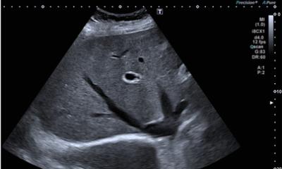
Intra-abdominal fat makes it harder for sound waves to penetrate through to the organs and obtain a good quality signal, which impacts image quality. As a result, ultrasound for larger patients hasn’t’ really been explored yet. It was a major concern for healthcare providers and the industry to develop appropriate solutions using the latest technology to provide obese patients with examinations using ultrasound, a non-ionising modality, Barbara Kraus, a radiographer at the University of Applied Sciences in Vienna, Austria, explained. "You must answer clinical questions in your report. Sometimes you had to write: 'you cannot present the lesion in an adequate form.' But you also have to achieve good resolution when the distance to the required depth of examination is greater," she said.
Companies are working on increasing depth of penetration to ensure that, where frequencies are required, the sufficient spatial resolution in the region is adequate to accommodate the larger field of view in abdominal ultrasound.
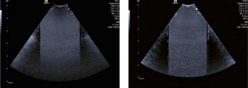
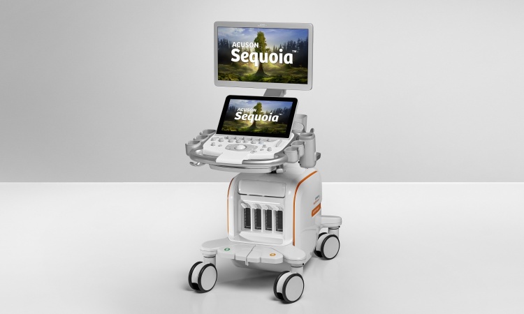
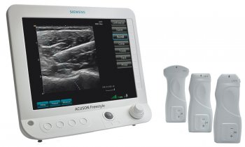
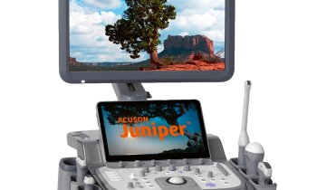
© 2020 Siemens Medical Solutions USA, Inc.
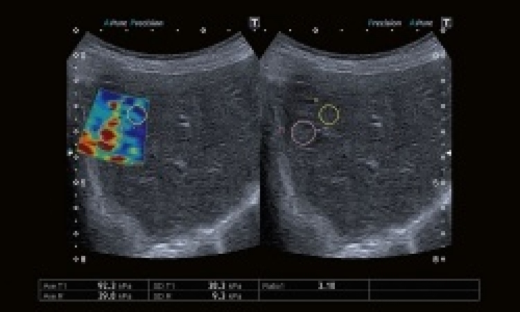
Non-focal imaging, another strategy developed by the industry, allows automatic adjustment of the focus position and saves time during the examination. Users can achieve higher frame rates and greater abdominal penetration in combination with a high focus rate. These new solutions offer not only higher spatial resolution, but also more time to focus on the patient, said Kraus, who presented the latest techniques available in the market during the session. "With deeper penetration, you get much more information from the signal coming back from the tissue. This was not possible with older devices. This technology takes much more information from the tissues and expands our field of investigation. Because we don't have to use the focus manually during the examination, we also save a lot of time that we can devote to our obese patients, who have the same rights to excellent imaging as any other patient," she concluded.
Profiles:
Jonathan McNulty, PhD, is Associate Dean for Graduate Taught Studies in the School of Medicine, University College Dublin, Ireland and President of the European Federation of Radiographers Societies.
Barbara Kraus, MSc, is a radiographer and researcher at the University of Applied Sciences in Vienna, Austria where she is responsible for Ultrasound Education in the Bachelor Degree Program of Radiological Technology, and Head of the Sonography Academic Course.
Reference
1 https://iopscience.iop.org/article/10.1088/1361-6498/aaf1dd/meta
17.12.2020