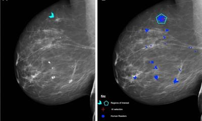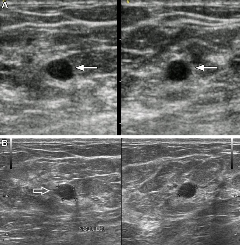
Image source: RSNA; from: Berg et al., Radiology 2023
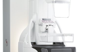
News • Using portable ultrasound imaging
AI helpful in triaging breast masses in low-resource areas
Artificial intelligence (AI) can analyze breast mass images from low-cost portable ultrasound machines and accurately identify cancer, according to a new study. This could prove useful for triage in low-resource settings.
The study was published in Radiology, a journal of the Radiological Society of North America (RSNA).
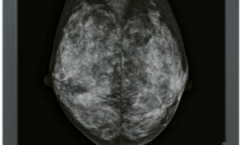
Breast lumps are often found accidentally, during breast self-exam or during a breast exam by a medical professional. Breast cancer screening can find cancers in the breast before the lump can be felt. While cancer screening has been the focus in Western countries, low- and middle-income countries often lack access to organized screening programs and technology.

In low- and middle-income countries, breast cancer most commonly presents as a palpable lump in the breast. Ultrasound can play a critical role in early detection, resulting in more effective, less invasive treatment and improved outcomes. “Women in low- and middle-income countries often cannot access breast health care for many months, even when they feel a lump in their breast that may be cancer,” said the study’s lead author Wendie A. Berg, M.D., Ph.D., professor of radiology at the University of Pittsburgh School of Medicine in Pittsburgh, Pennsylvania. “Our study looked at using AI to evaluate breast ultrasound images to distinguish suspicious breast masses needing urgent attention from those that were not cancer.”
For the multicenter study, participants with at least one palpable breast lump were enrolled in Jalisco, Mexico, from December 2017 through May 2021. Ultrasound images were obtained first with portable ultrasound at the site of the lump and adjacent tissue. Women were then imaged with standard-of-care ultrasound. Breast Imaging-Reporting and Data System (BI-RADS) assessments were performed by a radiologist.

Image source: RSNA; from: Berg et al., Radiology 2023
After exclusions, 758 masses in 300 women (mean age 50.0 years) were analyzed by the AI software as benign, probably benign, suspicious or malignant (cancerous). The mean patient age was 50.0 years (range 18-92), and the mean largest lesion diameter was 13 mm (range 2-54). Of 758 masses, 360 (47.5%) were palpable, and 56 (7.4%) malignant. AI correctly identified 96% and 98% of the women with cancer on the low-cost portable ultrasound and standard-of-care ultrasound images, respectively. Of the benign masses, 67% could have been appropriately triaged with standard-of-care ultrasound, and 38% with portable ultrasound.
Although specificity was less than with standard-of-care equipment, AI applied to portable breast ultrasound can potentially reduce about half of specialized hospital referrals in resource-limited regions.
Dr. Berg noted that the researchers did not train AI on images from the portable ultrasound. She also said that low-cost portable ultrasound technology has improved since the study was conducted. With better images and AI training, the researchers expect even better results in the future. “Our results show great promise for the use of AI and portable ultrasound in low-resource settings, including remote/underserved areas in the United States, to help improve breast health care,” Dr. Berg said. “In reducing the number of women with benign lumps who need to be seen in central facilities and potentially have a biopsy, health care resources can be better focused on women with cancer and reduce delays in diagnosis. This should improve access, health equity and outcomes for women.”
Source: Radiological Society of North America
03.05.2023