Image source: Adobe Stock/Mark Kostich
News • Study confirms association
CT scans in young people increase cancer risk
The EPI-CT study, which involved almost one million people, confirms that CT imaging, although largely beneficial, entails a small risk that needs to be minimised as much as possible.
A multinational study of almost one million individuals confirms a strong and clear association between exposure to radiation from CT scans in young people and an increased risk of blood cancers. This is the main conclusion of the analyses led by the Barcelona Institute for Global Health (ISGlobal), a centre supported by the ”la Caixa” Foundation, in the European-funded EPI-CT study. These results, published in Nature Medicine, highlight the importance of continuing to apply strict radiological protection measures, particularly in paediatric populations.
The benefits of computed tomography (CT) for imaging in patient management (including diagnostic efficacy, treatment planning and disease follow-up) are undisputed. However, the extensive use of this procedure in recent decades has raised concerns in the medical and scientific community about the potential cancer risks associated with exposure to ionising radiation, particularly in young patients. “The exposure associated with CT scans is considered low (less than 100 mGy), but it is still higher than for other diagnostic procedures,” says Elisabeth Cardis, Head of the Radiation Group at ISGlobal and senior author of the study. Previous studies have suggested an increased risk of cancer in in children exposed to CT scans, but they had several methodological limitations.
Recommended article

Article • Medical imaging
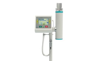
Computed tomography (CT)
Since its introduction in the 1970s, computed tomography has been a mainstay of radiology. Its overlay-free representation of body structures and the rapid image availability make CT indispensable in the diagnostic assessment of numerous diseases, especially in emergency medicine. Modern CT systems not only offer innovative procedures for better image quality, but also reduce radiation exposure.
To address these limitations, clinicians, epidemiologists and dosimetrists from nine European countries (Belgium, Denmark, France, Germany, Netherlands, Norway, Spain, Sweden, and UK) came together to conduct a multinational, European-funded study, EPI-CT, coordinated by the International Agency for Research on Cancer (IARC). “Implementing this large, multinational study was challenging – it involved extracting data from radiology records of 276 hospitals and linking them to population-based registries in nine countries, all while maintaining the confidentiality of the individuals’ data,” says Cardis.
The study analysed data from almost one million people, who underwent at least one CT scan before the age of 22. The dose of radiation delivered to the bone marrow, where blood cells are produced, was estimated for each person. By linking this information to national cancer registries, EPI-CT researchers were able to identify those who developed a blood cancer over time. Individuals were followed for an average of 7.8 years, although for those who had CT scans in the early years of the technology, researchers were able to monitor cancer incidence for more than 20 years after the first scan.
The procedure must be properly justified – taking into account possible alternatives - and optimised to ensure that doses are kept as low as possible while maintaining good image quality for the diagnosis
Elisabeth Cardis
The results show a clear association between the total radiation doses to the bone marrow from CT scans and the risk of developing both myeloid and lymphoid malignancies. A dose of 100 mGy multiplied the risk of developing a blood cancer by a factor of about 3. These results suggest that a typical scan today (with an average dose of about 8 mGy) increases the risk of developing these malignancies by about 16%. “In terms of absolute risk, this means that, for every 10,000 children who have a CT scan, we can expect to see about 1-2 cases of cancer in the 12 years following the examination,” says first author Magda Bosch de Basea, ISGlobal researcher at the time of the study.
The authors point out that more work is needed to ensure that doses and technical parameters are systematically and adequately collected in the clinics in real time to further improve risk estimates in the future.
Today, more than one million children in Europe undergo CT scans every year. Although radiation doses from CT scans have decreased substantially in recent years, the findings of this study underline the need to raise awareness among the medical community and to continue to apply strict radiation protection measures, especially in the youngest patients. “The procedure must be properly justified – taking into account possible alternatives - and optimised to ensure that doses are kept as low as possible while maintaining good image quality for the diagnosis,” Cardis explains.
Source: Barcelona Institute for Global Health
10.11.2023