Image source: MUSC
News • Mobile point-of-care imaging
An MRI-equipped ambulance: A game-changer for stroke care?
In the U.S., someone has a stroke every 40 seconds and dies from it every three minutes and 14 seconds, according to the Centers for Disease Control and Prevention. When it comes to stroke, experts echo the fact that time is brain. Faster treatment translates to better outcomes, and certain treatments, like the clot-busting drug tPA, have a strict time window for administration.
“The quicker that we can get the patient to treatment, the quicker we can have a good outcome,” said Dustin LeBlanc, M.D., director of Prehospital Medicine and associate chief medical officer for Emergency Management at the Medical University of South Carolina (MUSC). “But the big step in between – that is diagnostic studies.”
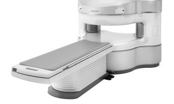
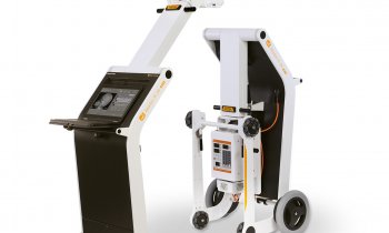
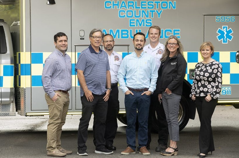
To accelerate diagnostic studies, a team of MUSC researchers, including LeBlanc, partnered with Charleston County Emergency Medical Services (EMS) to equip an ambulance with a portable MRI. The team was led by Donna Roberts, M.D., professor in the Department of Radiology and Radiological Sciences at MUSC and the deputy chief scientist for the International Space Station National Lab, and included Jillian Harvey, Ph.D., a professor in the College of Health Professions. Their findings, reported in the Journal of Stroke and Cerebrovascular Diseases, showcased the potential of the MRI-equipped ambulance.
An earlier trial in collaboration with Georgetown County EMS laid the foundation for this human trial by showing that images could be obtained in a moving ambulance on a standardized calibration model.
Recommended article
News • Neuroimaging
Stroke: MRI scan in an ambulance
Doctors work on initiatives to cut down the steps that need to happen between the time a stroke patient is wheeled through the ambulance bay until treatment can begin.
The project received funding from MUSC’s Blue Sky Award program, aimed at supporting high-risk research that tackles major medical and scientific challenges.

Image source: MUSC
Roberts thinks the new technology could be a game-changer for stroke care decision-making. “If you're somebody who could just receive tPA, you might go to a local hospital, while those who need to have advanced procedures, such as interventional neuroradiology, would go to a different hospital,” she said. “The imaging provided by the portable MRI scanner in the ambulance could help make that decision.”
Harvey thinks that it will also speed up stroke response times. “If we can get that information in transit and the decision process going before they even arrive at the hospital, then we can shorten the time to care and treatment,” she said.
Harvey is working with Kit Simpson, DrPh, also a professor in the College of Health Professions, to analyze state stroke data to see where MRI-equipped ambulances are most needed. “The real benefit may come in rural areas where patients may be hours or further away from an MRI,” said Harvey.
For LeBlanc, it’s all about how cutting-edge technology can transform and improve patient care. “If you think about where defibrillators were 50 years ago, they were hundreds of pounds, and it really took out-of-the-box thinking to imagine they could be portable. And now, they're public access points,” he said. “The MRI-equipped ambulance is just another example of technology helping us to develop ways to make things faster, lighter, smaller, more portable and to get it to the patient as quickly as possible.”
Ultimately, we need to stop thinking about telling patients to come to us. Instead, we need to be thinking about how we can take medicine to the patient
Donna Roberts
Ambulances equipped with CT scanners, known as mobile stroke units, are already used by some medical centers. However, they have limitations, including radiation exposure. The portable MRI has no radiation risk, and because it has a much lower magnetic field than traditional MRI, safety concerns regarding metal near the system are minimized, allowing operation of other medical or electrical equipment. Traditional MRI imaging can also identify stroke patients without a clear stroke timeline. Further research will determine if portable MRIs can achieve the same.
The research team worked with Charleston County EMS to secure a portable MRI in one of its ambulances and capture imaging while driving slowly around a parking lot, which had been blocked off with the help of MUSC Facilities. During the trial run, the research team obtained imaging of a healthy volunteer. Diagnostic-quality images were obtained and successfully transmitted to hospital radiologists for review. “This was really a team sport, with all aspects of MUSC and Charleston County EMS coming together for the mission of research,” said LeBlanc.
Despite these early promising results, more work needs to be done to see if diagnostic-quality images can be obtained when the ambulance is traveling at full speed. The team is also working with the Clemson School of Engineering to optimize the ergonomic fit of the MRI in the ambulance. With further technological advancements and clinical studies, MRI-equipped ambulances could become a game-changer in the field of emergency medicine, ensuring that stroke patients receive the best care possible, even before they reach the hospital. “Ultimately, we need to stop thinking about telling patients to come to us,” said Roberts. “Instead, we need to be thinking about how we can take medicine to the patient.”
Source: Medical University of South Carolina
02.11.2023