© vitanovski - stock.adobe.com
News • Noninvasive stratification
Multiplex PET helps doctors 'see' bowel cancer
Biopsies are currently used to diagnose bowel cancer but require an invasive procedure which offers risks, such as potential infection, and are unable to capture an entire picture of what is happening in a patient’s bowel.
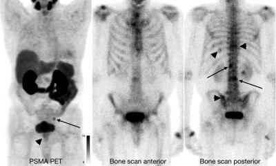
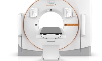
New research from the Cancer Research UK Scotland Institute and researchers at the University of Glasgow, published in Clinical Cancer Research, found that PET (Positron Emission Tomography) imaging could allow the entire bowel to be examined and tumours studied while inside the body, rather than examining tumour tissue once it has been removed.
PET scanning creates a three-dimensional picture of the inside of the body using labelled nutrients, or drugs known as a tracer, to show up areas where cells are more active than normal. The technology would allow bowel cancer patients to be matched to the best treatment for them, which is an area known as precision medicine, a growing area of oncology.
PET imaging offers a promising alternative, with the ability to survey the entire cancer landscape, allowing us to examine tumours in more detail while they are still growing
David Lewis
The team also believes multiple PET scans during a patient’s treatment could enable the progression of the cancer, and impact of treatment, to be effectively monitored. Dr David Lewis, of the Cancer Research UK Scotland Institute and the University of Glasgow, who led the research, said: “Precision medicine has the potential to revolutionise cancer diagnosis and treatment. However, the development of accurate, informative, and patient-friendly diagnostic techniques is crucial for its success. PET imaging offers a promising alternative, with the ability to survey the entire cancer landscape, allowing us to examine tumours in more detail while they are still growing.”
Each year around 4,000 people are diagnosed with bowel cancer in Scotland. With around 1,800 Scots sadly losing their lives to the disease annually, the search for new and better treatments is vital.
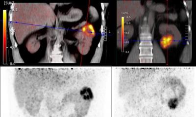
The research team used existing genetic data on bowel cancer to identify different tumour characteristics using PET imaging. They also discovered that multiple tracer PET scanning, which uses different PET tracers, rather than just one, was able to distinguish between different types of bowel cancer in mice based on their genes. This would allow a more personalised approach for patients as treatments could be tailored to their particular cancer.
Patients can have different mutations in their bowel cancer, and mutations in genes such as KRAS, APC and TGFB have different image signatures. The team believes PET imaging could identify the type of bowel cancer a patient has, by matching the image signature with the mutations, therefore allowing the patient to access the best treatment for their individual disease more quickly.
Dr Catherine Elliott, Director of Research at Cancer Research UK, said: “These findings by the team at the Cancer Research UK Scotland Institute and University of Glasgow offer an exciting opportunity to revolutionise the way we diagnose and monitor bowel cancer without invasive surgery, reducing the risk and improving outcomes for patients. PET imaging is a crucial tool in our future approach to diagnosing this disease which affects so many people in Scotland.”
Source: University of Glasgow
20.03.2024