assessment of gastrointestinal tumours and found PET/MRI to be more effective in detecting lesions and metastases
Article • Hybrid imaging
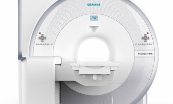
PET/MRI leads hybrid imaging
Hybrid imaging is still a leading topic in radiology – underlined by the 14 related sessions held during the 29th European Congress of Radiology (ECR 2017) held in Vienna, this March. Those sessions focused on the combination of radiological and nuclear medical imaging procedures that aim to visualise morphology as well as function, structure and metabolism of an organ or region of interest.
Report: Michael Krassnitzer

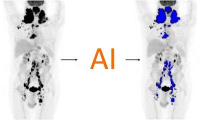
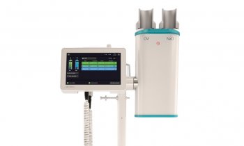
Currently, many radiologists consider positron emission tomography plus magnetic resonance imaging – PET/MRI – to be the most promising candidate in hybrid imaging.
Among them, Professor Katrine Åhlström Riklund has no doubt: ‘PET/MRI is going to be huge.’ The professor, who is deputy director of the Department of Nuclear Research, medical director of the Nuclear Medicine Department and director of the Medical School at Umeå University in Sweden, pointed out a current study of 2,300 patients that has shown PET/MRI and PET/CT to yield equally good results in oncological imaging (Spick C et al., J Nucl Med. 2016). Today, 18F-FDG PET/CT is the gold standard any imaging participant has to meet.
In the Hybrid Imaging in Oncology several smaller studies were presented which shared one major insight. While PET/MRI has been proven equal, or even slightly superior, to other procedures it has failed to create the dramatic breakthrough that has been predicted for a number of years.
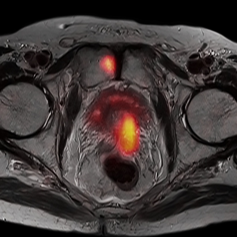
The very first study presented showed rather disappointing results. In pre-operative staging of surgical therapy of cervical cancer hybrid imaging did not offer meaningful additional information. ‘When determining the Peritoneal Cancer Index, PET/MRI does not show any advantages over MRI alone,’ Dr Montserrat Alemany Ripoll, Medical Director of the Department of Radiology at University Hospital Uppsala (Sweden) concluded.
However, a German study found PET/MRI to be superior to MRI in pre-operative staging of cervical cancer. ‘The results demonstrate the usefulness of 18F-FDG PET data as a valuable additive to MRI alone for more accurate assessment of nodal or distant metastatic spread in patients with primary cervical cancer,’ said Dr Johannes Grüneisen, junior physician at the Institute of Diagnostic and Interventional Radiology at University Hospital Essen.
At the same institute the diagnostic performance of PET/MRI and MRI alone was assessed in recurrent soft tissue carcinoma. Hybrid imaging results were positive, as the junior physician Dr Youssef Erfanian pointed out. ’18F-FDG PET/MRI displays superior detection accuracy compared to routine MRI follow-up examinations.’
A Turkish study compared PET/MRI and non-contrast enhanced PET in the assessment of gastrointestinal tumours and found PET/MRI to be more effective in detecting lesions and metastases. ‘We could detect significantly more lesions with the hybrid procedure,’ Dr Filiz Çelebi of the Department of Radiology at Bilim University, Istanbul, explained.
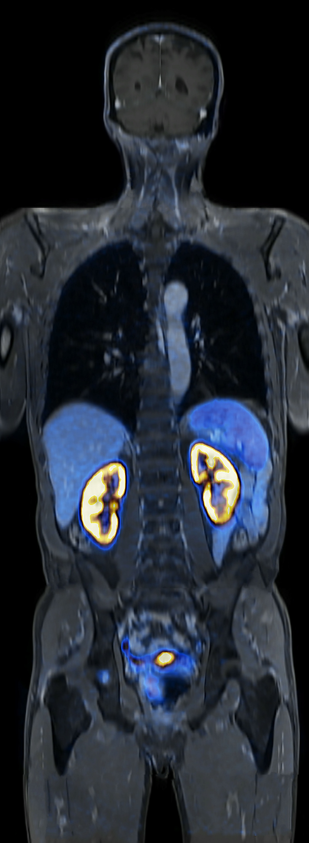
Even results that do not evidence the superiority of PET/MRI can be described positively as a comparative study on PET/MRT and PET/CT in the staging of neuroendocrine tumours shows. ‘68Ga-DOTATOC PET/MRI provides an equivalent diagnostic performance for whole-body staging of patients with neuroendocrine tumours compared with PET/CT,’ said Dr Lino Sawicki, researcher at the Institute of Diagnostic and Interventional Radiology at Düsseldorf University Hospital.
Profile:
Professor Katrine Riklund is an expert in radiology and nuclear medicine. She is deputy director of the Department of Nuclear Research, medical director of the Nuclear Medicine Department and director of the Medical School at Umeå University in Sweden. She was also President of the ECR 2016.
03.11.2017