Article • Underrated technique
Pitfalls in pelvic CT imaging
Computed tomography (CT) plays an increasingly important role in assessing pelvic disease, particularly when patients present with acute abdominal pain. In addition, radiomic approaches on CT are being developed to increase the characterisation of ovarian cancer for optimising treatment planning.
Report: Mark Nicholls

The subject, and wider role of CT in pelvic conditions, will be the focus of a presentation – “Pelvic disorders: is there a role for CT?” – at the Garmisch-Partenkirchen CT Symposium from Professor Andrea Rockall of Imperial College London.
She said: “Patients presenting with pelvic discomfort frequently undergo ultrasound as the first line imaging investigation, particular in women suspected of pelvic disease, with MRI being used for specific indications or problem solving. However, CT is widely used in the acute setting, when patients present with peritonitis or so-called acute ‘surgical’ abdomen and knowledge of underlying acute diagnoses in the pelvis is essential.”
Differentiation difficulties
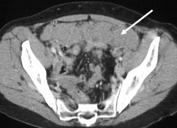
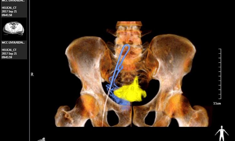
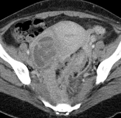
While it is important to be aware of the potential pitfalls, as well as incidental findings that may be encountered, she said that CT remains the standard of care for some non-acute pelvic disease evaluation, including staging of ovarian cancer, though other modalities such as FDG-PET/CT and MRI are being increasingly used in this context. During the session she will highlight what some of the pitfalls are – such as the example of disseminated ovarian cancer on CT being difficult to differentiate from bowel - and how to spot incidental findings on pelvic CT, as well as detailing ovarian cancer imaging on CT and the use of radiomic approaches.
The main pelvic disorders are inflammatory conditions and cancer with CT offering the significant benefit to the clinician/radiologist in that it is quick and usually available. However, Professor Rockall, who is Clinical Chair of Radiology at Imperial College London and Honorary Consultant Radiologist at Imperial College Healthcare NHS Trust and at The Royal Marsden Hospital, added: “With CT, gynaecologic disease of the uterus and ovaries can be difficult to interpret. Currently, there is significant interest in evaluating ovarian masses on CT using a radiomic approach and the current findings in radiomic approaches in ovarian cancer will be discussed. “Radiomic approaches in ovarian cancer may allow triage into optimal treatment strategy, leading to better patient outcomes.”
Also useful for post-operative assessment
Another learning objective to be outlined by Professor Rockall is to ensure delegates can be familiar with the appearances of acute pelvic disease on CT, including gynaecological and non-gynaecological pathology, with a number of examples and case studies discussed during the session. Professor Rockall, who will focus on the role of CT in female patients, said: “Patients attending accident and emergency departments with abdominal or pelvic pain frequently have CT when interpretation of the pelvic findings is so important, and not always easy.”
MRI remains the standard modality for local staging of cervical cancer - and endometrial cancer in UK – as it allows much better delineation of the gynae soft tissues, though she acknowledges that it is not always required or available, particularly in an emergency setting, for example. She added that CT is also useful in post-operative assessment where there may be a suspected complication.
Professor Rockall is currently the Chief Investigator of the CRUK MAPPING trial in cervix and endometrial cancer and three NIHR trials MALIBO, MALIMAR (machine learning studies) and MROC (a multi-centre UK trial evaluating multi-parametric MRI in suspected or confirmed ovarian cancer).
Profile:
Professor Andrea Rockall is Clinical Chair of Radiology at Imperial College London and Honorary Consultant Radiologist at Imperial College Healthcare NHS Trust and at The Royal Marsden Hospital. Her special interests are in genitourinary cancer, image-based clinical trials, functional imaging in response assessment and machine-learning applications in radiology.
Presentation:
Freitag, 24.01.2020, 11.10-11.30 Uhr
Pelvic disorders: Is there a role for CT?
Andrea Rockall (London)
Session: Onkologie
24.01.2020