Source: University of California, Los Angeles (UCLA), Health Sciences
News • Imaging
PSMA PET/CT beats standard scan at prostate cancer detecting
Prostate-specific membrane antigen imaging should be the positron emission tomography imaging agent of choice for men who have a prostate cancer recurrence after radical prostatectomy, which needs to be detected and located. For these men, it should become the standard of care, new research shows.
After prostate cancer surgery, it is important that patients are routinely screened for a recurrence of cancer. This screening is done with a blood test that checks for the presence of the prostate-specific antigen, or PSA. If this test comes back positive, it means that there is a recurrence of cancer. But this test doesn’t indicate where the cancer is located.
Doctors know that the location of recurring prostate cancer after surgery is often in the prostate fossa, the area where the prostate was before it was removed. But in some cases the cancer can be located outside of the prostate fossa, such as in the lymph nodes or in the bones.
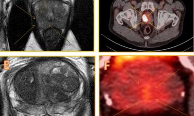
An imaging technique called fluciclovine positron emission tomography, or Axumin PET/CT, is currently the standard of care in the United States for finding the location of the prostate cancer recurrence. However, another new imaging method exists that is called prostate-specific membrane antigen imaging, or PSMA PET/CT, that can also be used for the same purposes. This scan is currently investigational and has not yet been approved by the FDA.
Researchers from the UCLA Jonsson Comprehensive Cancer Center undertook a head-to-head comparison of these two imaging techniques and have concluded that prostate-specific membrane antigen imaging is more effective in detecting the location of the prostate cancer recurrence.
Background
As men age their prostate glands produce increasing levels of prostate-specific antigens. Levels above a certain threshold can be a sign of cancer. For men who have been treated for prostate cancer, a high level of prostate-specific antigens can signal a recurrence of the disease, commonly referred to as biochemical recurrence. In the case of prostate surgical removal, the presence of prostate-specific antigens in the blood means that some prostate cancer cells remain in the body even after prostate removal.
Physicians try to determine the exact location of the biochemical recurrence by using a fluciclovine PET/CT scan. With this technique, doctors inject the men with fluciclovine, which mimics amino acids that doctors can track because they are radioactive. Since cancer cells consume more amino acids than regular cells, the fluciclovine imaging technique is able to pinpoint the location of the cancer by tracking the cells that are consuming more of the fluciclovine.
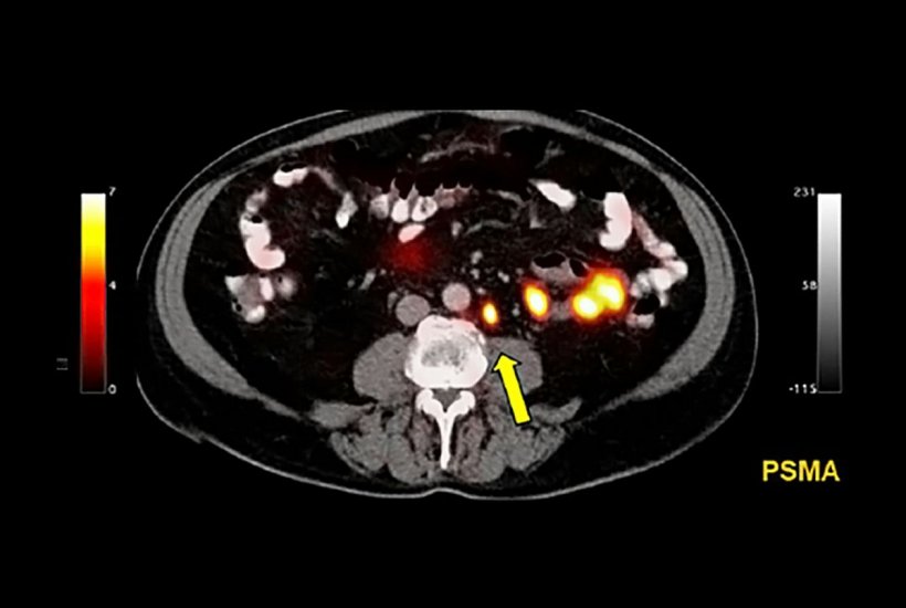
In contrast, the prostate-specific membrane antigen imaging technique tracks the antigens’ protein expression levels. Since prostate cancer cells overexpress a high level of prostate-specific membrane antigens in comparison to normal cells, this imaging method can determine the location of the cancer cells by tracking these cell surface proteins.
Research
To compare the effectiveness of these two imaging methods, UCLA researchers studied 50 patients. All of the men in the clinical trial had previously had a radical prostatectomy where surgeons removed the prostate gland and surrounding tissue due to cancer. They all had a biochemical recurrence of prostate cancer with low prostate-specific antigen levels which indicated that their cancer recurrence was at an early stage.
The men each received a fluciclovine scan as part of their post-surgical recurrence standard-of-care treatment. The men also agreed to receive an additional prostate-specific membrane antigen imaging scan within 15 days before or after their fluciclovine scan.
For the purpose of the study, UCLA researchers removed each individual’s personal information from the scan images. These images were sent to three independent physician readers. The scans were read three times by each reader. The readers were unaware of the findings of the others.
The results showed that the prostate-specific membrane antigen imaging was able to detect the recurrence of prostate cancer in 56 percent of the scans. In comparison, the fluciclovine scan was able to detect prostate cancer in 26 percent of the scans.
Source: University of California, Los Angeles (UCLA), Health Sciences
04.08.2019