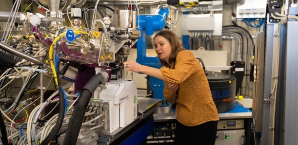
Image source: Lund University/Agata Garpenlind
News • Updating treatments
Sugar and fat can make cancer cells harder to kill
In their quest to find new and better methods to make cancer cells more susceptible to treatment, Karin Lindkvist and her research group at Lund University in Sweden are looking into the world of molecules, using the X-rays at the MAX IV laboratory.
Author: Agata Garpenlind (Lund University)
The researchers believe that limiting the cells' access to sugar will make cancer cells more sensitive to treatment. Many of the cancer treatments used today were established in the 1980s. In order to decrease mortality further, there is an urgent need for new ways to treat cancer. Researchers around the globe have taken on this challenge, including Karin Lindkvist and her team in Lund. “We are combining structural biology with cell biology to find new molecular mechanisms that facilitate the development of new treatments for cancer”, says Karin Lindkvist.
Targeting the glucose uptake of the cancer cells may represent a new treatment for overcoming chemo resistance
Karin Lindkvist
The blood cancer Acute Myeloid Leukemia (AML) is the most common form of leukemia in adults and the second most common in children. Despite the introduction of new drugs, the overall cure rates remain around 20 percent in adults and 65 percent in children.
Emerging literature suggests that obesity is a risk factor for cancer. Obesity further reduces leukemia survival, and increases the risk of treatment-related complications, suggesting that leukemia cells residing in adipose tissue (fat cells) may be more resistant to treatment and therefore contribute to disease persistence.
However, the nature of the interplay between leukemia cells and fat tissue is poorly understood. “Leukemic stem cells have been shown to utilize the fat tissue microenvironment to support their metabolism, that is, they use the fat cell as energy and this helps them survive chemotherapy. We have preliminary data suggesting that leukemic cells become more sensitive to chemotherapy if we block their glucose uptake. Therefore, targeting the glucose uptake of the cancer cells may represent a new treatment for overcoming chemo resistance”, explains Professor Lindkvist.
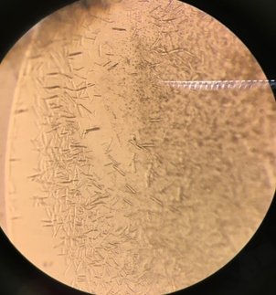
Image source: Lund University
To perform this type of research, large amounts of human protein is required. Since one research obstacle is obtaining sufficient amounts of human protein, the researchers use the gene coding for the specific protein, for example a glucose (sugar) transporter, to duplicate it. Next the protein must be well-packed into a crystal in order for the researchers to study the details of the protein structure and how the cancer drug is binding to it. “We're interested in blocking the sugar uptake of cancer cells, in order to improve cancer treatment. But in order to do so we need to know how the proteins look and thus how cancer drugs target them”, says Karin Lindkvist.
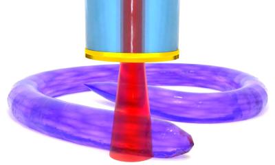
In their research the group utilizes the beamline facility at the MAXIV laboratory. “A regular microscope isn't powerful enough for looking closer at this type of proteins. So instead we expose the proteins to X-rays from the beamline. The light is dispersed and then we collect the reflections and calculate what the protein looked like in the crystal”, says Karin Lindkvist. Soon a new beamline called MicroMAX will be built at MAX IV. This infrastructure addition will facilitate the collection of research data for Karin Lindkvist in that the crystals used can be much smaller.
Source: Lund University
04.02.2020