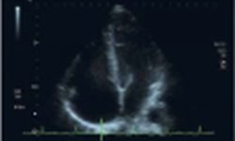
Video • Transthoracic ultrasound localization microscopy
Super-resolution imaging of microscopic heart vessels
A new imaging technique tested on patients could improve the evaluation of cardiac conditions and undiagnosed chest pain.
Researchers from Imperial College London’s Department of Bioengineering and Faculty of Medicine worked alongside academics from University College London to produce sub-millimetre resolution images of cardiac micro-vessels. The non-invasive new imaging technique was tested on four human patients.
Existing imaging technologies can visualise large vessels on the heart’s surface. However, this new technique could allow scientists to study the physiology of the heart in more detail by imaging smaller micro-vessels within the heart muscle. This research, published in Nature Biomedical Engineering, could help clinicians better understand the role such vessels play in cardiovascular diseases such as microvascular coronary disease and cardiomyopathies, as well as undiagnosed chest pains.
This has opened up a wide range of opportunities to study heart physiology and observe different diseases and conditions non-invasively and safely
Mengxing Tang
Professor Mengxing Tang, from the Department of Bioengineering at Imperial College London, and the corresponding author of the research, said: “Visualising cardiac vessels is crucial for managing cardiovascular diseases, but there is a lack of understanding of how the blood flows within the small vessels of the heart. Our study images these vessels non-invasively in the highest resolution which, following further research, could help clinicians to manage these diseases.”
The heart relies on efficient blood flow to be able to pump blood around the body, supplying tissues with oxygen while removing carbon dioxide and waste. However, damaged heart vessels can result in abnormal blood flow, potentially causing tissue injury leading to heart failure.
The scientists tested the imaging technique on four patients with hypertrophic cardiomyopathy (HCM), a condition that makes the walls of the heart chamber thicker with abnormal tissue and reduces the amount of blood pumped in and out. They used ultrasounds and microbubbles (small, gas-filled bubbles used to differentiate between internal structures in medical imaging) to image the microvascular structure and flow dynamics of the patients’ hearts in super-resolution. The data was collected at St Bartholomew’s Hospital in London. The size of the micro-vessels, combined with the fast movements of the heart, made imaging them challenging, especially at resolutions under a millimetre.
The researcher’s technique could potentially help to evaluate different cardiac conditions. For example, clinicians could use the technique to visualise structural abnormalities in patients with microvascular coronary disease and cardiomyopathies, making it easier to diagnose and treat, thus improving health outcomes.
Professor Tang said: “This is the first time we demonstrated it is possible to image these vessels in such resolution, which has never been done before in humans. This has opened up a wide range of opportunities to study heart physiology and observe different diseases and conditions non-invasively and safely.”
Recommended article

Article • Research, diagnostics, therapies
Focus on Cardiology
Arrhythmias, valve defects, heart attacks: Cardiologists face a wide range of disorders and diseases. Equally diverse are diagnostics and therapeutic options. Find out what the field has to offer.
Co-author and cardiologist Professor Roxy Senior, from the National Heart and Lung Institute at Imperial College London, said, “For the first time this technique allows direct visualisation of the very small heart muscle vessels which when diseased give rise to chest pain which can be not only debilitating but may also lead to death. Because at present these vessels can be assessed only by indirect means the condition can be misdiagnosed.”
The research is still in its early stages, despite initial success, so more studies will be needed on more patients to better understand the clinical and research value of the technique. Further research may also lead to improvements in image quality. Professor Tang said: “We are optimistic about what our new technique could bring to cardiovascular patient healthcare.”
Professor Tang is also exploring the potential use of super-resolution ultrasound technologies for evaluating a range of other diseases, working with oncologists, cardiologists, radiologists, breast surgeons and other clinicians. He said: “This far-reaching research would never happen without collaboration between interdisciplinary teams of engineering and clinical sciences researchers.”
Source: © Imperial College London; by Helena Kudiabor
08.05.2024