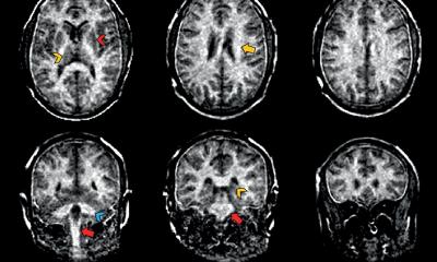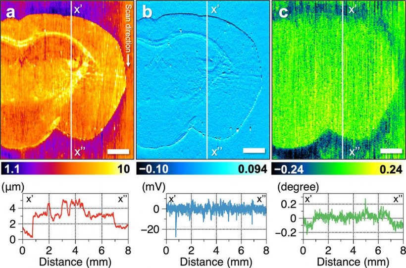
Image source: Atsuka et al., Analytical Chemistry 2021 (CC BY-NC-ND 4.0)
News • Neuroimaging research
Using mass spectrometry to diagnose brain diseases
To quickly and correctly diagnose diseases, medical professionals depend on identifying what biochemicals are present in tissue sections, where the biomolecules are, and at what concentrations. Mass spectrometry imaging—which can identify multiple biochemicals in a single experiment—is a promising technique to achieve this. However, the stability of biomolecular sampling needs improvement to obtain the chemical distribution information with high spatial resolution.
In a recent study published in Analytical Chemistry, researchers from Osaka University used mass spectrometry to image the distribution of fat molecules in mouse brain tissue. They acquired data at a spatial resolution of 6.5 micrometers, enabling analysis on a cellular level.
The researchers used a very small capillary to gently extract lipid molecules from a tissue section, and a carefully designed setup for fine 3D directional control. Although biological tissue may often seem smooth to the naked eye, on an ultrasmall scale it's rather rough. The ability to account for this ultrasmall-scale roughness is central to obtaining reproducible biochemical data at high spatial resolution. "In our experiments, the probe's vibration amplitude is constant even when the sample height changes," says Yoichi Otsuka, first author. "We can also measure changes in sample height up to 20 micrometers, and it can be increased up to 180 micrometers."
Principle component analysis helped us integrate our wide-ranging data. For example, we could assign the classes of lipids that are primarily present in the cortex and brainstem
Takuya Matsumoto
The researchers' first experiments were to measure irregular distributions of molecules across an uneven surface: microwells filled with various concentrations of a dye. The measured concentrations correlated with the known concentrations, and the measured surface topography was close to the actual microwell diameter. Experiments with mouse brain sections yielded a multi-dimensional data of multiple molecules such as the distribution of certain hexosylceramides—lipids that are important in aging. "Principle component analysis helped us integrate our wide-ranging data," explains Takuya Matsumoto, senior author. "For example, we could assign the classes of lipids that are primarily present in the cortex and brainstem."
Correlating such data with disease progression will require further study and perhaps additional development of the researchers' biomolecule extraction setup. The researchers anticipate that their approach will be useful for quantitatively imaging the myriad neural networks in brain tissue. Ultimately, they hope to help medical practitioners reliably diagnose diseases such as brain cancer in a tissue section with the support of molecular information in high spatial resolution.
Source: Osaka University
20.01.2021